Penetration
 View Larger
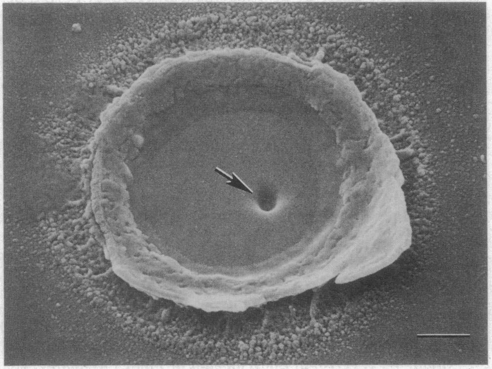
FIG. 2. Scanning electron micrograph documenting surface penetration of Mylar sample 5. A site of penetration was exposed by sonicating the Mylar, shearing the attached appressorium, leaving a ring of attached cell wall surrounding the appressorium pore (8). Within the pore a hole in the Mylar is visible (arrow), produced by a penetration peg (see ref. 11) that was removed during sonication. (Bar = 1 Am.). From: Howard, R.J., Ferrari, M.A., Roach, D.H. and Money, N.P. 1991. Penetration of hard substrates by a fungus employing enormous turgor pressures. Proc. Natl. Acad. Sci. 88:11281-11284.


